Surgical Correction of Truncus Arteriosus (Type II) in a Neonate
Truncus arteriosus (TA) represents 1-2% of congenital heart defects in liveborn infants. Based on an estimated incidence of congenital heart disease of 6-8 per 1,000 liveborn children, truncus arteriosus occurs in approximately 5-15 of 100,000 live births(1) and TA Type II obviously can be seen even rarer.
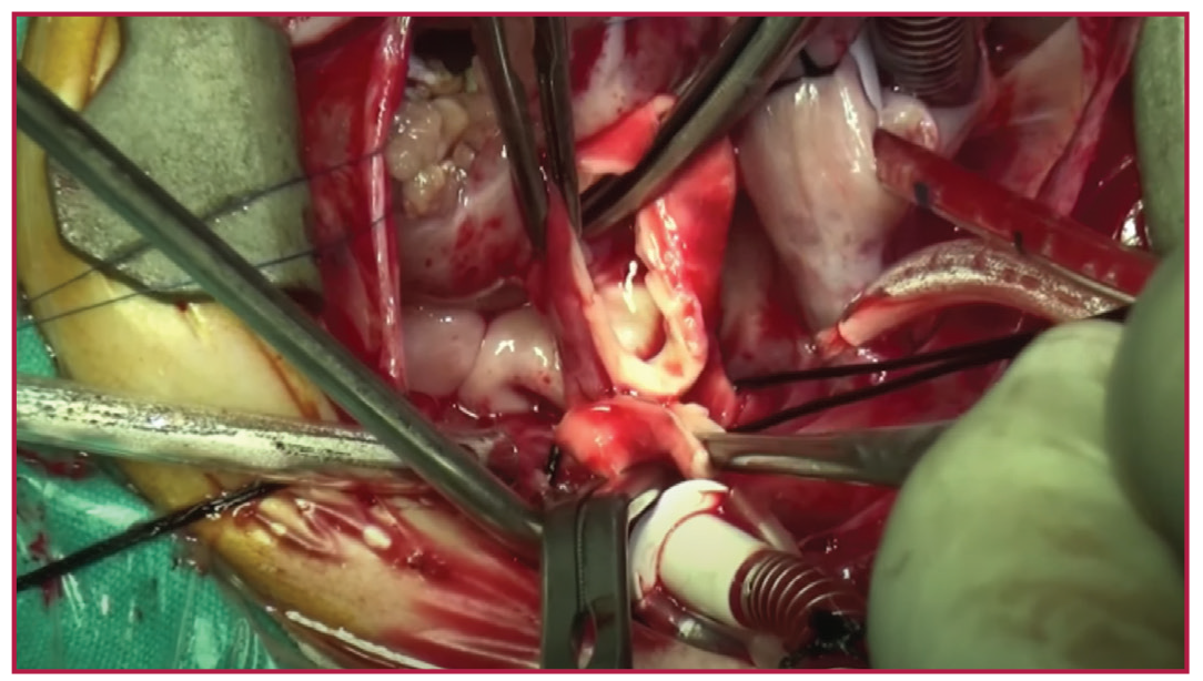
We present here a surgical repair video of a 45 days old baby with Type II TA. She had a usual large conal ventricular septal defect (VSD), mild aortic regurgitation and bilateral posterior orifices of both pulmonary arteries from the TA.
The VSD was closed with a large heterologous pericardial patch. The bilateral pulmonary artery that was taking off from the aorta, was carefully excised as a button. A 16 mm Contegra Medtronic bovine jugular vein conduit was anastomosed between the right ventricle outflow track and pulmonary artery button.
In addition to repair of TA with an external conduit, a piece of the left manubrium sternum was excised as an important preventive method for early post-operative period, in order to prevent the conduit to be compressed by sternum. This technique is almost a routine procedure that I perform to prevent the conduit compression in my practice.
The patient was discharged without any complication.
References
McElhinney, Doff B. “Truncus Arteriosus.” Background, Pathophysiology, Etiology, 31 Dec. 2019. https://emedicine.medscape.com/article/892489-overview?src.